Insights
The Importance of visualizing complex details in your sample and using them in their full context
In materials science, it's vital to consider both narrow and broader context to avoid incorrect assumptions. Suppose the cross section prepared by FIB is too small or taken from a defect-free area? Will our conclusion be that the material we produce is of flawless quality? And what if 3D tomography is performed on a small volume and at shallow depth, revealing small grains overall?
Read our paper and learn more about why contextual data is essential for accurate materials investigation.
UHR Field Free FIB-SEM Imaging
All details from all samples
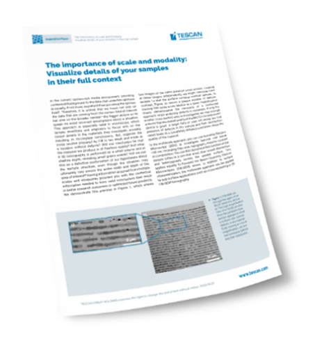
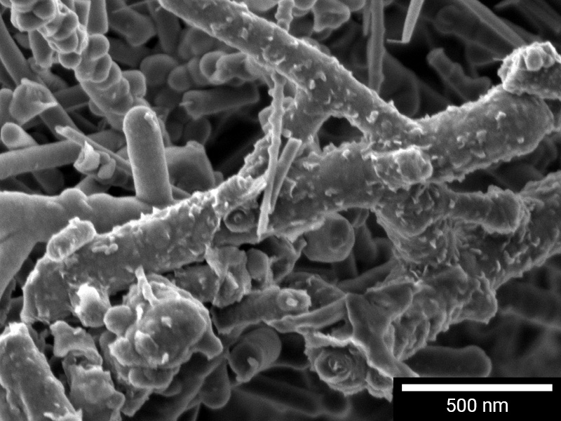
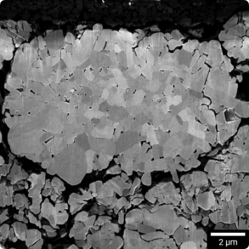
With field-free UHR technology, you can quickly and easily visualize and analyze topography, phase contrast, and more, regardless of your sample's characteristics. Whether your specimen is metallic, magnetic, non-conductive, charging, or beam-sensitive, our ultra-high-resolution imaging performance delivers the low keV and high surface sensitivity needed for accurate analysis.

Visualize the truth. Consistently and with maximum contrast
Contrast is key to a true understanding of surface topography and material composition. TESCAN-designed Backscattered Electron (BSE) detectors are made for today’s materials, with attention to not only conventional samples, but also beam-sensitive, emissive, or heated samples.

Widest selection of BSE contrast methods
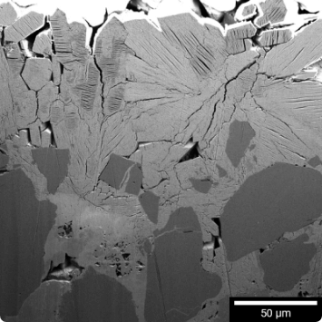
As the diversity in analyzed specimens grows, different BSE contrast methods are required to properly describe phases of the investigated specimens. For this reason, it is crucial to select the appropriate BSE detector for the kind of information you need to collect from your sample.


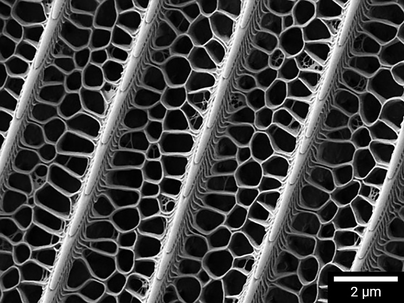
Macro. Micro. Nano.
Start at 2x for navigation, then zoom to your feature of interest, collecting images at any point. Wide Field optics, UHR SEM and FIB work together to deliver multi-scale data, at and below the surface.

TESCAN Wide Field™ mode
TESCAN Wide Field Optics™ mode is standard on all TESCAN SEMs and FIB-SEMs and provides the most intuitive navigation experience by displaying the industry’s widest undistorted field of view in the live SEM window.


3D Multimodal Characterization with Unmatched Speed and Precision
Meet Mistral™ - the most precise and powerful plasma FIB on the market. Engineered for applications demanding precision, like TEM sample preparation. Mistral™ offers optimized resolution and the highest beam current at the same time. Whether you’re working with high or low currents, Mistral™ delivers the best resolution across all settings.
Challenging materials? No problem.
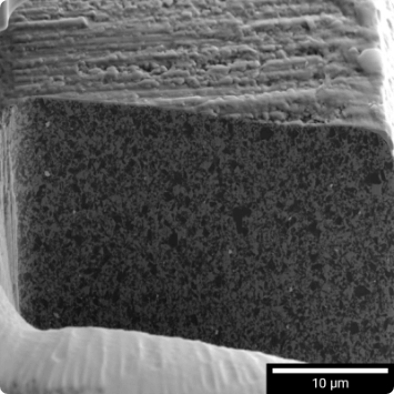
Hard and soft material mixes, excessive topography or preferential sample orientation can create poor FIB-cut surface quality. TESCAN’s Rocking Stage and True X-Sectioning enable fast cross-sectioning and 3D analysis without compromising throughput or surface quality.


TESCAN Rocking Stage
TESCAN Rocking Stage is fully compatible with TESCAN hardware and software to create an integrated solution for more efficient workflow execution and improved milling speed, accuracy and quality.


TESCAN True X-Sectioning
TESCAN’s TRUE X-Sectioning employs a masking principle for large-scale (above 200x200 µm²) 3D characterization tasks, where conventional deposition methods are significantly slower and impractical. This approach allows for higher beam currents, speeding up the entire characterization process.


3D Nanotomography. Effortless, but mighty.
Specify the volume of interest. Define your acquisition parameters or follow our step-by-step guided workflows, then begin data collection. TESCAN’s Essence Tomography module makes it fast and straightforward for any operator to successfully perform 3D sample analysis.
And TESCAN’s own powerful 3D rendering engine delivers high-quality visualizations with several viewing options and exports comprehensive rendered animations.

Versatility Meets Precision
Discover the TESCAN Essence Multimodal FIB-SEM Tomography, seamlessly integrated into Amber X 2’s Essence™ graphical user interface. This advanced tomography module supports a wide range of imaging and analytical detectors, offering unmatched versatility and precision for your imaging needs.


3D EDS and EBSD
Always Accurate. Always Fast.
Understand relationships between structure, composition, and crystallography – even in 3D. Our patented static setup for FIB-SEM slicing and 3D tomographic data acquisition ensures large volume 3D EBSD analysis that is both reliable and fast. Experience unparalleled efficiency and accuracy in every slice and dataset.

TESCAN 3D Viewer software
TESCAN’s 3D Viewer module is easy to learn, so both new and experienced users can produce complex 3D visualizations quickly. A step-by-step wizard guides the operator through the import, alignment, and pre-processing steps in their correct sequence, resulting in complete, detailed output for 3D visualization.


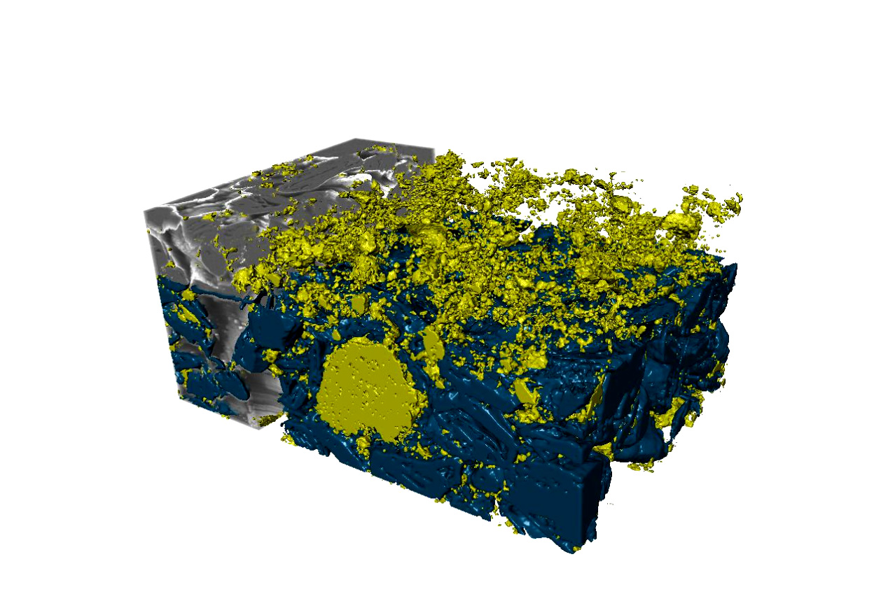
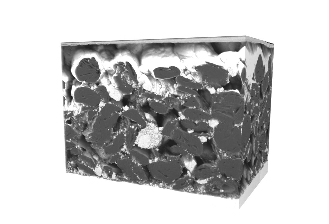
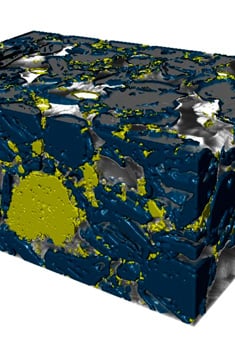
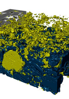
3D Investigation of New Battery Technology Material Structure and Chemistry
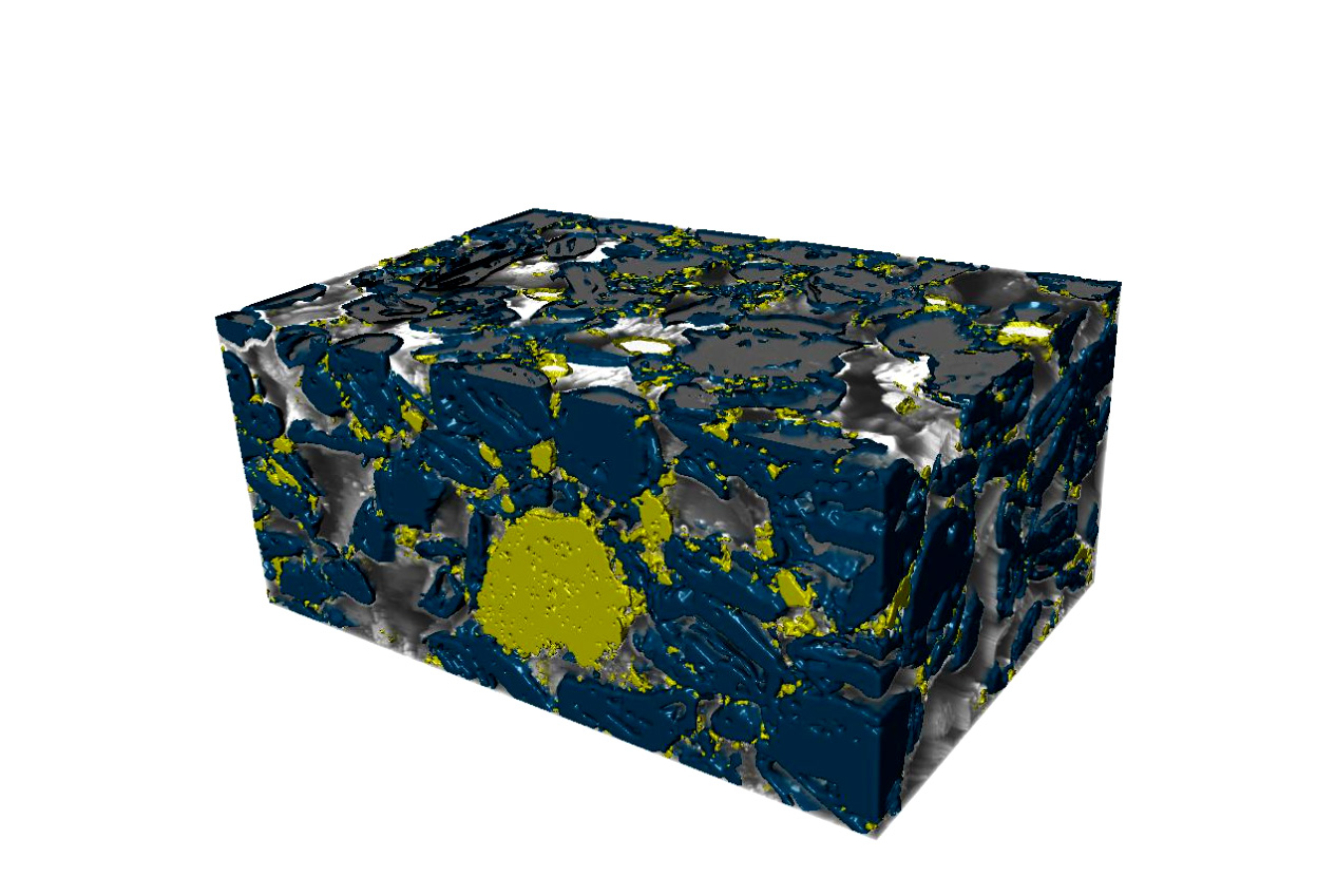
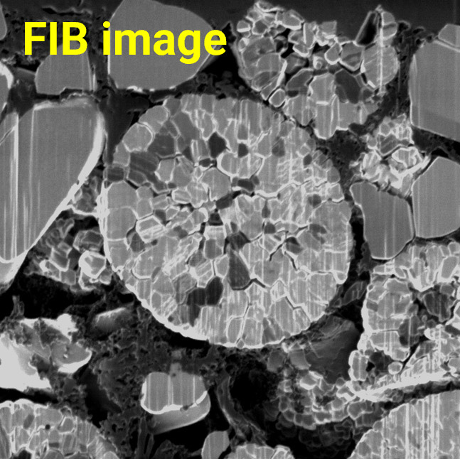
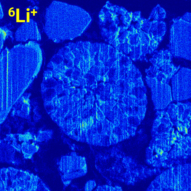
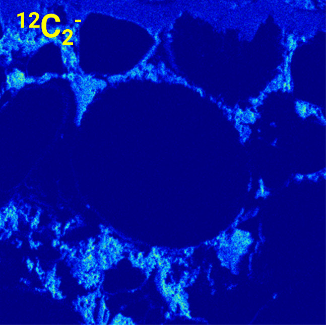
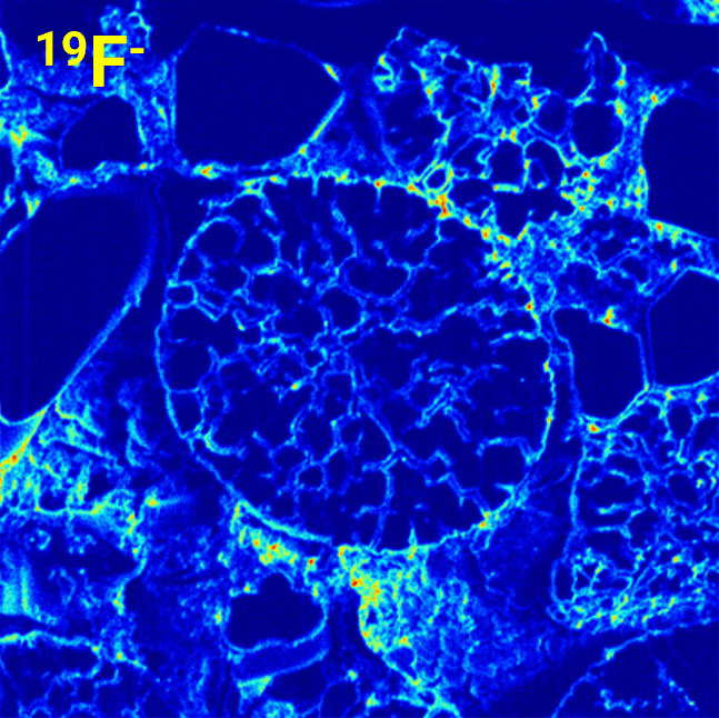
Understanding the structure and chemical composition of battery materials is pivotal to determining the behavior and stability of new battery chemistries during the cycling process. Grasping the complex relationship between local morphology and chemical state, such as particle, void, lithium, and binder distribution within electrodes, is vital for performance optimization and degradation resistance for these advanced battery materials.
By utilizing 3D FIB-SEM tomography combined with ToF-SIMS analysis, we can conduct detailed investigations of the material composition in new battery technologies. This 3D approach delivers more precise volumetric statistical data than traditional ToF-SIMS depth profiling. Moreover, 3D ToF-SIMS tomography aids in pinpointing contaminants, vulnerabilities, and chemical inconsistencies within the battery components.

3D ToF-SIMS: Beyond Battery Research
Use the power of 3D FIB-SEM tomography combined with ToF-SIMS analysis for a thorough examination of emerging materials. This method offers more accurate volumetric statistical information compared to conventional 2D ToF-SIMS depth profiling. 3D ToF-SIMS tomography is not only crucial for advancing battery technology but also holds promise for other areas of Materials Science, enabling precise mapping of specific elements, isotopes, or trace elements.


Embrace the Lightest Elements with Integrated ToF-SIMS
Analyze light elements and characterize the chemistry of samples containing Li, C, H, O or LiP with <50 nm spatial resolution at the ppm concentration level. This is made possible using integrated TESCAN ToF-SIMS for 2D and 3D chemical analysis and visualization.


Unlock Faster, Cleaner Results in Carbon-Based Materials
Combine the TESCAN Mistral™ column with the OptiGIS O2 oxygen injection module to transform your FIB-SEM workflow. This integration:
-
Speeds up material removal
-
Enhances milling precision
-
Reduces contamination for cleaner cross-sections
Achieve Precision in Silicon and Aluminium Milling
TESCAN OptiGIS O2 gives you control—slowing down milling when needed for high-fidelity shape and depth accuracy.
-
Targeted oxygen delivery enhances material removal dynamics
-
Achieve efficient, predictable outcomes with minimal tuning
-
Maintain consistent, repeatable results in even the most demanding applications

Have no fear. Move those samples.
Essence™ Collision model is always on guard, preventing any hardware collision inside the closed FIB-SEM chamber during stage movement, sample tilting and detector insertion.

TESCAN Essence™ 3D Collision Model
The Essence™ 3D Collision Model replicates the chamber interior and stage and detector motion. In doing so, it creates a virtual model that prevents collisions and helps users to adjust hardware positions for collision-free movements.