
Exploring Pore-Scale Fluid Flow Using Dynamic Micro-CT
The study of fluid migration through porous media is essential for understanding various natural and industrial processes. These include the...
By Wesley de Boever, Product Marketing Manager for MicroCT, TESCAN
“Versatility” is an often mentioned and highlighted feature for many micro-CT systems. However, since most micro-CT users rely on their system for standard sample analysis, it is a feature that is often neglected and not taken into consideration when purchasing a new micro-CT system.
Indeed, when only standard micro-CT scans are performed, all you will ever need in terms of system and sample movement is a rotation of the sample, a translation of the sample between source and detector – to change the field of view and resolution – and preferably a vertical movement of the sample – to scan the sample at different heights or perform stacked scans.
Having only a few extra degrees of freedom can already greatly improve the user experience and results of a micro-CT scanner. The most obvious example is the ability to change the basic geometry of the scanner by having a variable source-to-detector distance (SDD). This implies that the detector can be shifted closer to the X-ray source to increase the X-ray flux at the detector, and therefore reduce the scan speed. The effect of this movement is greater than one would expect, since the amount of X-ray flux versus distance follows an inverse square law (figure 1), meaning that doubling SDD divides the flux by four.
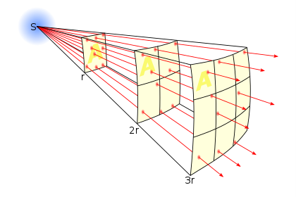
Figure 1: Inverse square law: flux decreases with distance squared - source: Wikipedia.
But having a variable SDD also works the other way around. Being able to move the detector far away – up to a staggering 2 meters in the TESCAN UniTOM XL – means high resolution can be maintained, even when in-situ devices limit the freedom to come very close to the source with the sample. As an example, in the UniTOM XL, a voxel size of 5 µm can be obtained for objects up to about 15 cm in diameter.
There are many other movements inside a CT system that can be beneficial. A translation of the sample stage perpendicular to the source-detector axis can be used to move tall samples out of the way to acquire open-beam (flat field) images to measure the intensity I0 when no sample is in the beam path. Moving the detector from side-to-side enables the acquisition of tiled (wide-field) datasets, doubling the horizontal field of view of a scan. Having additional motorized detector stages allows users to automatically switch between different detectors, even extending the basic imaging capabilities of a micro-CT scanner with different modalities.
Of course, scanner versatility can only be exploited to its full capacity if the operating software can make the best use of it. TESCAN‘s AcquilaTM acquisition software is designed to make full use of all degrees of freedom – up to no less than 10 – of the TESCAN UniTOM XL and HR systems. The intuitive scripting language enables individual control of all motors controlling the source, detector, and rotation stage―a concept nicely illustrated in this open access paper by our customers at the University of Antwerp, using the TESCAN UniTOM XL. A good application of this full system flexibility is Extended Cone Tomography™, where the source and detector need to be positioned at different heights, beneficial for the scanning of large and flat samples. As illustrated in figure 2, scanning this type of sample in a standard CT scan can cause artefacts due to the long path the X-rays need to travel through the material, especially when dealing with high-Z materials such as steel. Extended Cone Tomography™ tilts the optical axis of the CT system, getting a shorter beam path through the sample, and a clear, artefact-free image through flat samples.
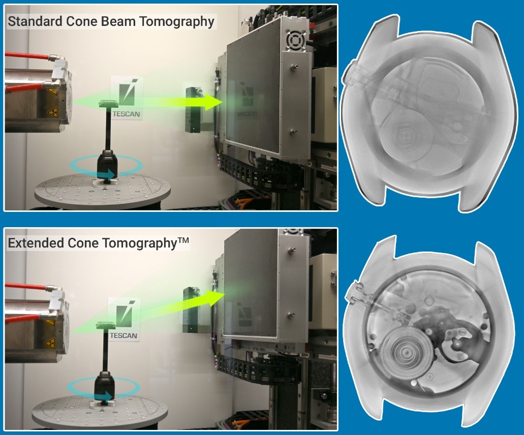
Figure 2: Improved imaging of flat samples using TESCAN Extended Cone TomographyTM.
For more information on TESCAN UniTOM HR and XL, please visit our website.

Wesley De Boever
Product marketing Manager for TESCAN micro-CT
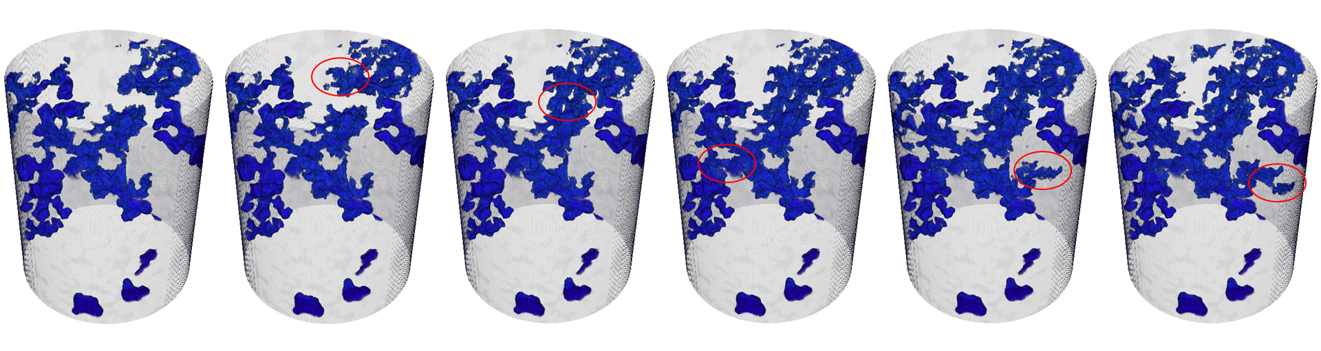
The study of fluid migration through porous media is essential for understanding various natural and industrial processes. These include the...
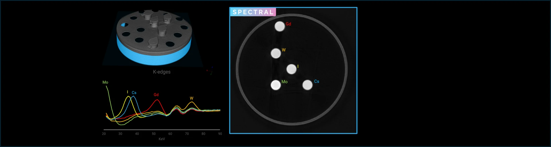
Spectral micro-CT imaging is a unique imaging modality that allows absolute element identification through K-edge imaging. This is extremely useful...
Spectral CT can be applied for detailed analysis of soil samples to identify both the components of the soil as contaminants present in the...